Diagnosis
Cranial Cruciate Ligament Rupture and Disease
Cranial Cruciate Ligament (CCL) rupture is the most common cause of rear limb lameness in dogs. The CCL is the equivalent of the Anterior Collateral Ligament (ACL) in humans.
It is one of two ligaments that cross inside the stifle joint. The stifle is the veterinary term for the canine knee. These ligaments prevent the femur and tibia from sliding back and forth on each other.
The biomechanical stress, together with inflammatory or genetic factors, causes fraying of the ligament, resulting in a partial or complete tear.
Cause of Cranial Cruciate Ruptures
The cause of cruciate ruptures is most often multifactorial and degenerative. The name for this condition is canine Cranial Cruciate Disease (CCLD). Factors that exacerbate the disease and biomechanics are obesity, poor nutrition, joint inflammation, limb deformities, insufficient or erratic exercise, and genetics.
If you suspect that your companion has suffered a CCL injury, please familiarize yourself with the common signs and symptoms and contact your veterinarian to schedule a physical exam.
Signs of a Cranial Cruciate Rupture:
- Sudden lameness on a rear limb to a degree where weight can not be borne
- A history of mild limb lameness that comes and goes before a sudden worsening of symptoms
Signs of Lameness:
- Pain
- Decreased range of motion
- Muscle atrophy (loss of muscle mass)
- Abnormal posture when standing, getting up, lying down or sitting
- Abnormal gait when walking, trotting, climbing stairs or turning
- Nervous system signs — confusion, trembling, etc.
- Swelling and inflammation
- Grating or grinding joint movement
While CCL ruptures are the most common cause for hind limb lameness, the entire limb, and pet should be assessed to rule out hip disease, ankle arthritis, neurological factors, common comorbidities, and secondary injuries due to a CCL rupture.
A frequent secondary injury of a CCL tear is patella luxation and instability.
The best thing a pet owner can do if they suspect their dog is experiencing any form of hind limb lameness is to contact a veterinary specialist and schedule a physical exam, as symptoms may worsen without treatment.
Consequences of a CCL Rupture and Disease:
- Ligament deterioration releases a combination of inflammatory factors from the ligament
- Increasing instability and inflammation of the joint from the weakened ligament causes arthritis to develop quickly within the joint.
- Chronic instability increases the risk of damage to the meniscus- a structure crucial for joint health.
Weight-bearing studies that use force plate analysis have shown that dogs with a damaged CCL bear only 20 to 30% of the weight they would typically place on the affected leg.
As a result, pets will compensate and start shifting their weight onto their healthy limb. Placing more pressure and stress on the opposite limb increases the possibility of damaging the other cruciate ligament and stifle. It is common for an animal to rupture the CCL in both stifles.
Every time the pet bears weight on the affected leg, the femur slides down the tibial plateau with nothing to halt its movement. This sliding action damages a cartilage seal in the joint called the meniscus. Once the meniscus tears, arthritic change accelerates, and pain worsens.
Treatment Options
Due to the frequency of cranial cruciate injuries and a need for a solution, the veterinary profession has focused tremendous energy on this problem. Resulting in a variety of treatment options ranging from very conservative treatment options to invasive surgical procedures.
Pain Management
The most conservative treatment option is pain management and the prescribing of pain drugs. Although pain management drugs may help the dog feel better and cope with a sore knee, they DO NOT alter the progression of the disease.
Surgical Treatments
Surgical treatments for the cruciate disease are generally divided into two categories: lateral suture stabilization and geometry modifying osteotomies.
Lateral suture stabilization procedures use durable braided materials to hold the tibia in place relative to the femur. The suture material links the femur to the tibia by fixing the ends with bone anchors or tunnels with buttons. These techniques provide a short-term constraint that facilitates the development of joint fibrosis, which increases stability. Small and toy breeds are considered the best candidates for these techniques.
Dogs that are medium, large, and active breeds are sometimes not the best candidates for this technique as they are more likely to overwhelm the suture and require further treatment.
Geometry modifying osteotomies seeks to neutralize the joint force on the torn CCL by cutting and rotating the top of the tibia or cutting a segment of the tibia and advancing it forward.
Treatment Option #1: TPLO Surgery
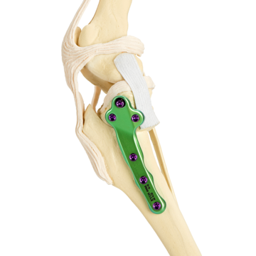
 Tibial Plateau Leveling Osteotomy (TPLO) surgery is the first broadly accepted geometry modifying orthopedic procedure introduced by Dr. Barclay Slocum. The TPLO surgery involves a curved cut bisecting the proximal end (top) of the tibia separates the tibial plateau from the rest of the bone. The tibial plateau is then rotated to a position that resists the slipping of the femur over the tibia. The final step is that the bone segments are secured by a plate and locking screws.
KYON ALPS® TPLO implant gives animals advantages during and after the operation. The contoured shape and consistent locking mechanism make surgical application convenient and routine. Biocompatible titanium materials and design protect against infection, which remains the most common complication associated with conventional stainless steel locking plates.
Treatment Option #2: TTA Surgery
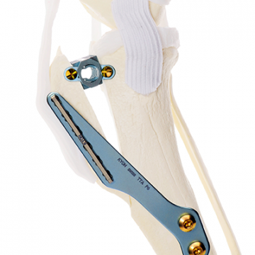
 Tibial Tuberosity Advancement (TTA) surgery can achieve the same objective as TPLO surgery, with less disruption to the contact mechanics of the joint when treating the most common cause of lameness in dogs. KYON founder, Slobodan Tepic. Then, a professor of surgery at the University of Zürich, Pierre Montavon, developed the TTA implant and procedure as a less aggressive method to counteract the joint force. A straight cut separates a segment of bone that contains the attachment of the patellar ligament. This segment is then advanced, using the patellar tendon to act as a surrogate CCL. The advancement is then secured by a novel tension band plate, fork, and cage implant.
KYON TTA implant and surgery is clinically proven to be successful. The procedure has been used in over 150,000 cases by more than 1,000 surgeons worldwide. The broad range in available cage sizes, shape, and biocompatible titanium reduces post-surgical complications and accelerates recovery.
Treatment Option #3: TTA-2 Surgery
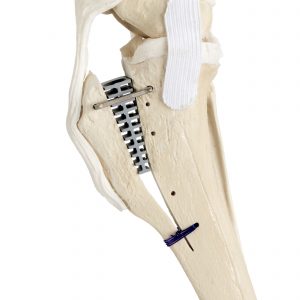
 Tibial Tuberosity Advancement-2 (TTA-2) surgery was developed by KYON to improve upon the original TTA. The TTA-2 system is an even less invasive and simplified surgical technique that repairs the ligament.
The KYON TTA-2 system differs from the original TTA because 1) it requires smaller and fewer holes in the tibia and, 2) it requires stapling, eliminating the need for screws. Most importantly, the TTA-2 implant and procedure are clinically proven to be successful. With over four years of a clinical study where 700 cases performed, and more than 6,000 procedures performed since its launch.
Secondary Treatment(s)
Secondary treatments are treatments to correct secondary injuries. As mentioned previously, a CCL rupture often results in patella luxation and instability, where further treatment may be needed.
Patellar Groove Replacement (PGR)
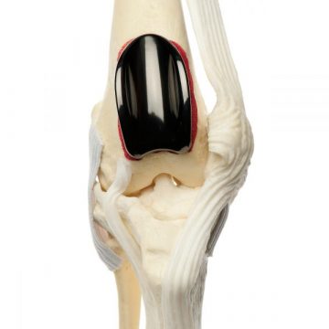
 TTA and TPLO surgeries can correct the alignment of the patella on its own for some patients. However, when the condition is more severe, a partial joint replacement like Patellar Groove Replacement (PGR) can be combined with TTA or TPLO surgery.
The Patellar Groove Replacement implant is a unique solution to address the condition of a patellar luxation where the kneecap dislocates from its location in the patellar groove. It is the first and only device on the market to restore a functional patellar groove and provides immediate stability to the patella.
Recommendations
Following these three suggestions will help ensure your dog lives a happy and healthy life:
#1: Know the signs and symptoms of CCL rupture and disease
#2: If showing symptoms, have your dog tested for a CCL rupture and disease.
#3: DON’T WAIT, seek treatment if your dog has a CCL rupture/tear.
It is tempting to wait as long as possible before committing to a treatment option. However, as the injury or disease progresses, drastic changes to the joint and decreases a dog’s function and quality of life. Treating the diseased joint sooner-than-later will extend the active-life of your dog, saving years of mild and moderate lameness.